SCABIES: MYTHS TO DISPEL
Please note: to enlarge photo, click on it and return with browser back arrow
00 INSTRUMENTAL PROCEDURES
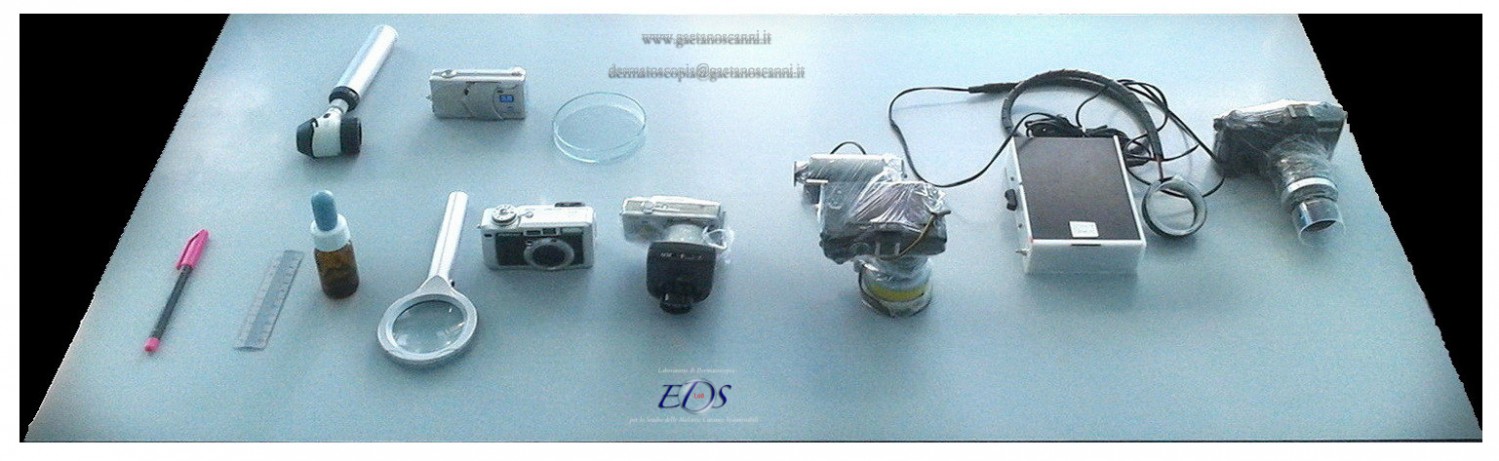
Fig 01. Some instrument used for an entodermoscopic examination. Particular attention was paid to the protection of tools with disposable transparent film.
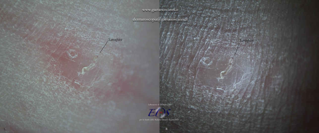
Fig 02. Contactless dermoscopy (d-DS). Comparison between two different types of lighting. On the left an image with polarized light, on the right the same lesion with non-polarized tangential light.
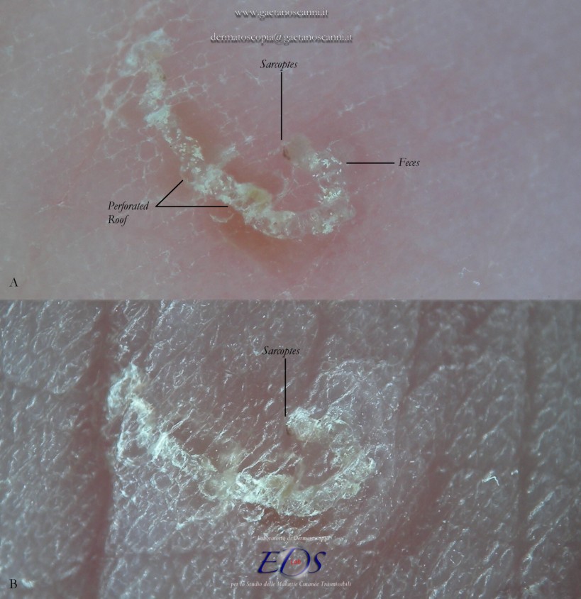
Fig 03. Contactless dermoscopy (d-DS). Above polarized light image allows to recognize the mite and feces inside the tunnel. Under the same image with tangential light that enhances the surface texture of the whole skin, highlighting the discontinuity of the gallery roof.
01 GLOBAL DESCRIPTORS
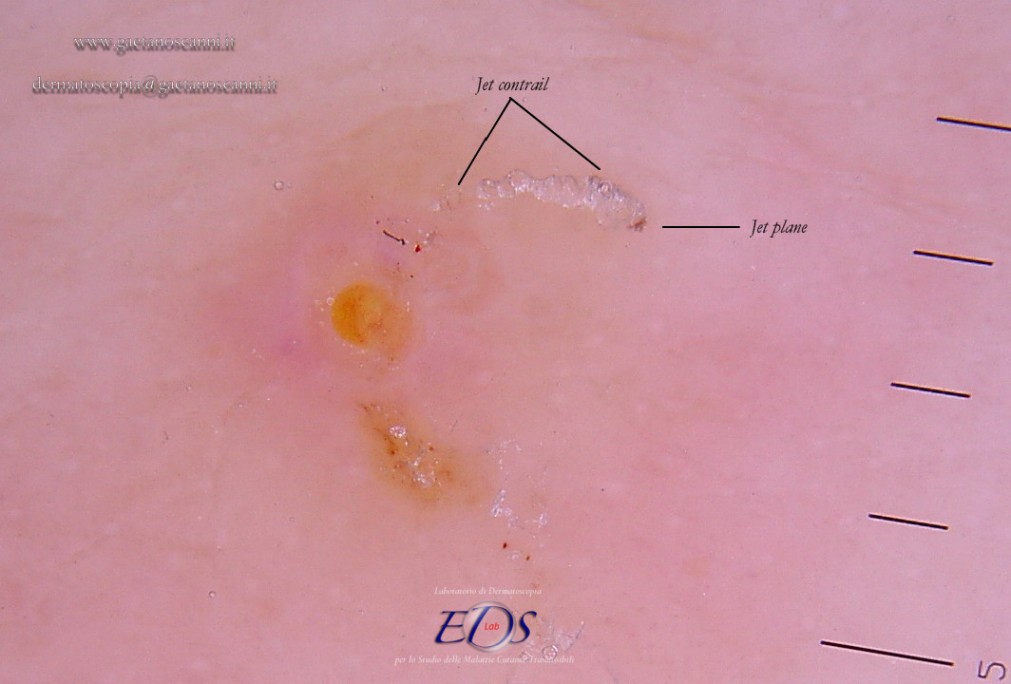
Figure 04. Contact dermoscopy (w-DS). The front of the parasite is recognized as a dark triangle resembling a delta wing of a jet. Immediately after, the tunnel produces a series of micro bubbles that remain trapped under the glass plate of the dermatoscope simulating the jet contrail.
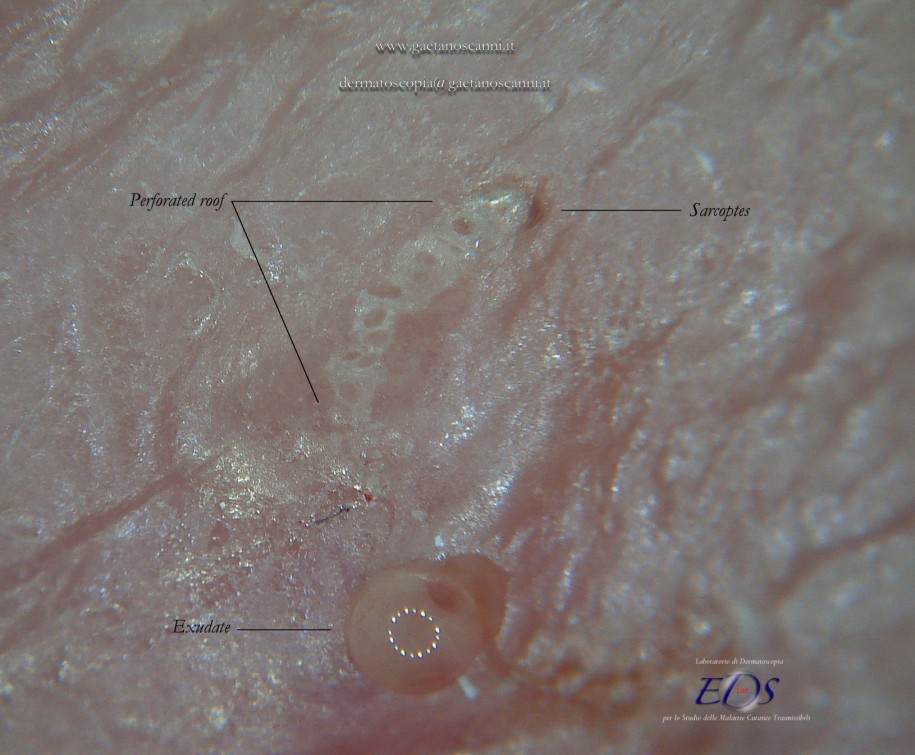
Fig. 05 Contactless dermoscopy (d-DS). The initial part of the tunnel where the mite resides has an intact roof. It is important to note that the gallery has instead a roof interrupted by holes from which air bubbles come out when switching to the w-DS (plate + liquid medium).
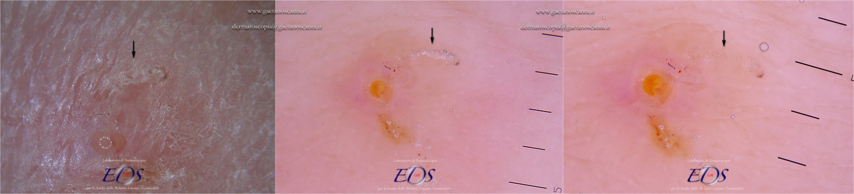
Fig 06. Contactless versus contact dermoscopy of the same Mite-Gallery Unit. In d-DS the roof of the tunnel appears interrupted by several holes that allow the air to exit (Left). The first description of an MGU resembling a white jet contrail was determined by the trapped air between the tunnel and the glass plate of w-DS (Center). After having discontinued dermoscope contact, the gallery disappears as all of air bubbles escape and the liquid medium passes inside it (Right).
02 LOCAL DESCRIPTORS
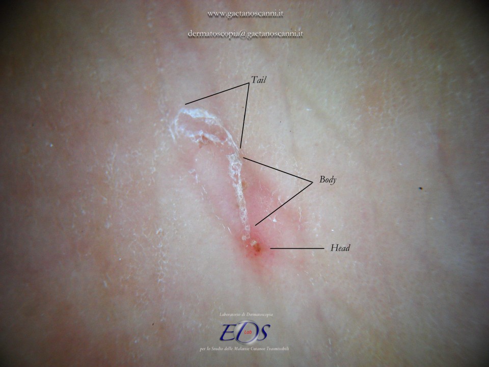
Fig 07. The Mite Gallery Unit under contactless dermoscopy (d-DS). The Mite Gallery Unit (MGU) structure can be divided into three parts. The Head hosting the mite, the Body which represents what clinically is defined as the burrow containing eggs and feces of the parasite and the Tail at the end of structure, which is an incomplete gallery because without roof but made of keratinic collarettes visible only in d-DS. An erythema is present in the background around and immediately behind the mite.
HEAD MGU
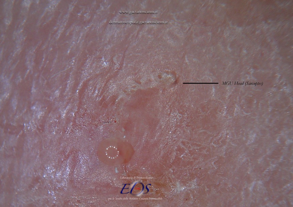
Fig. 08. Contactless dermoscopy (d-DS). The initial part of the MGU is the head. This part of the gallery houses the mite which is recognized by a small triangular area with the vertex oriented towards healthy skin where it will proceed. The head of gallery has a roof intact.
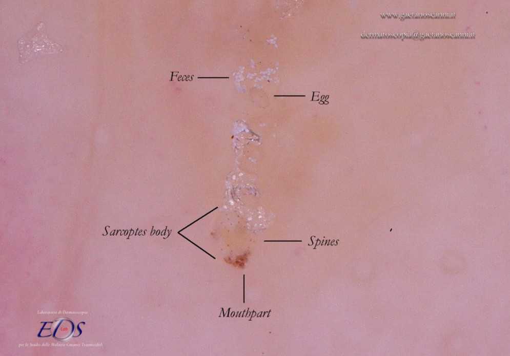
Fig 09. Enhanced contact dermoscopy of an MGU in w-DS mode. The Body of the mite. In addition to refraction of its anterior part, Sarcoptes scabiei shows an opalescent body with several scattered little dark dots (ladybug sign). These formations correspond to the "bristles" on the body that guarantee, among other functions, the adherence of the mite within the tunnel.
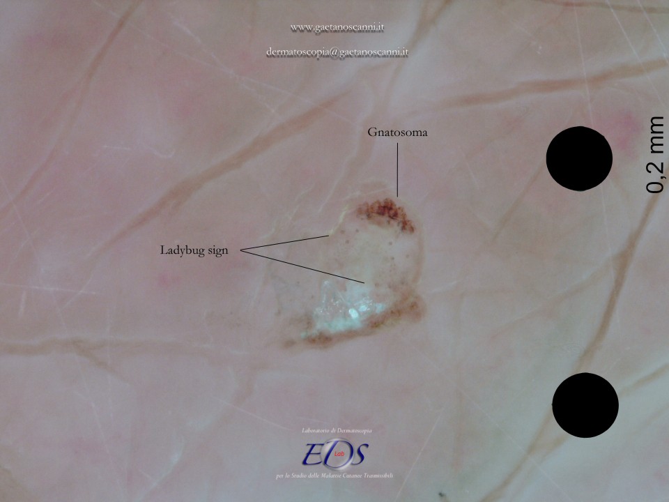
Fig 10 Enhanced contact dermoscopy (w-DS). The body of the mite has a slight opalescence which may be detected only at higher magnifications. The front part corresponds to the gnatosoma and the first pair of legs. The body of the mite is marked by little dark dots that correspond to the bristles of the back (mechano-sensory receptors) that give rise to the dermoscopic sign of the “ladybug”.
BODY MGU
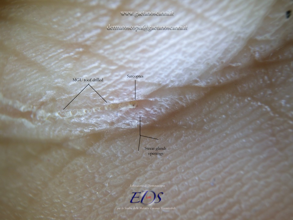
Fig 11. Contactless dermoscopy (d-DS) with lateral light. The body of the MGU corresponds to the main part of the tunnel on the bottom of which the mite eggs are glued. The roof of the tunnel is clearly interrupted by holes from which the larvae are supposed to come out but which could also be the result of the detachment of the sweat gland openings.
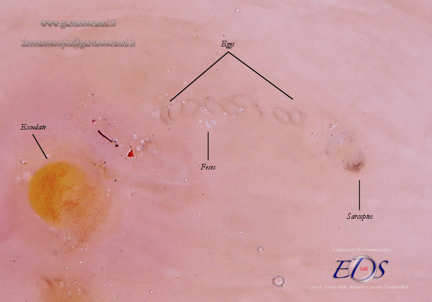
Fig 12. Enhanced contact dermoscopy of an MGU in w-DS mode. Content of the gallery. The absence of bubbles and the penetration of the liquid film in the tunnel, makes the roof transparent and allows to glimpse a row of eggs otherwise not accessible. The white dots are the feces of the mite. A serum-purulent drop is located on the left.
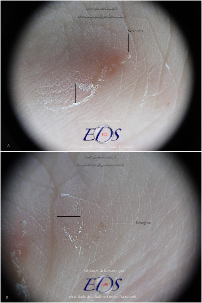
Fig 13. Contactless dermoscopy (d-DS). The rear of the MGU is no longer a gallery because it lacks the roof. In its place one can recognize two keratin edges that move away from each other. This phenomenon could be attributable to the turnover of the outer layers of the skin that replace the older part of the tunnel with new skin.
ATYPICAL MGU
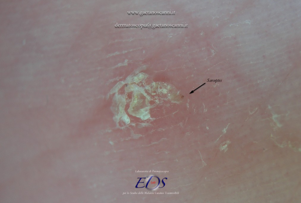
Fig 14. Contactless dermoscopy (d-DS) of an atypical MGU. The refractive part of the mite (arrow) is evident, behind which there is an aggregate of scales rather than an ordinary gallery.
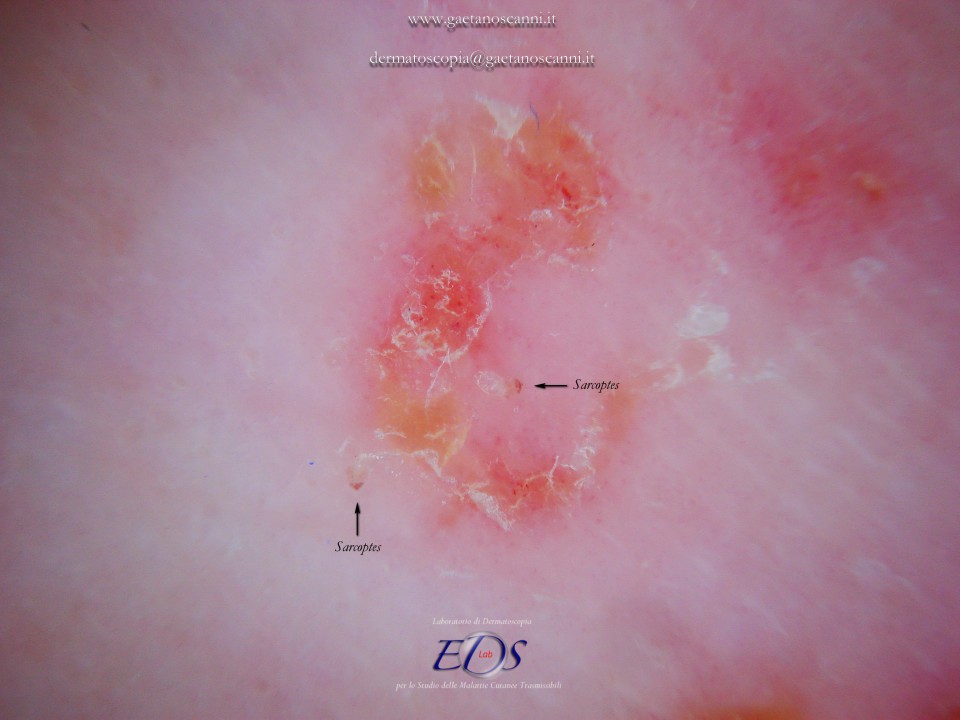
Fig 15. Contacless dermoscopy (d-DS) with polarized light of an atypical MGU. Two mites (arrows) can be recognized behind which there is no tunnel but an erythematous area with polycyclic borders.
03 MGU COMPLICATED BY PHLOGOSIS OR HYPERKERATOSIS
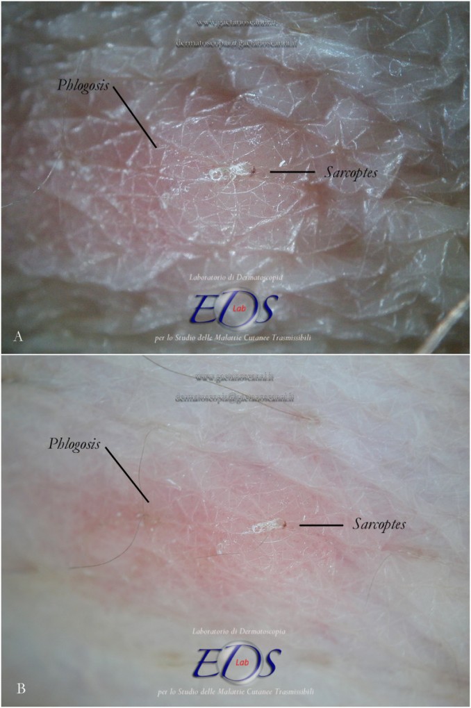
Fig 16. Contactless dermatoscopy (d-Ds). (A) tangential light, (B) polarized light. The images show an MGU in which can be appreciated the delta wing sign produced by the mite and the following body of the gallery. A pink area is visible around MGU which corresponds to the inflammatory response that the mite products induce in the host.
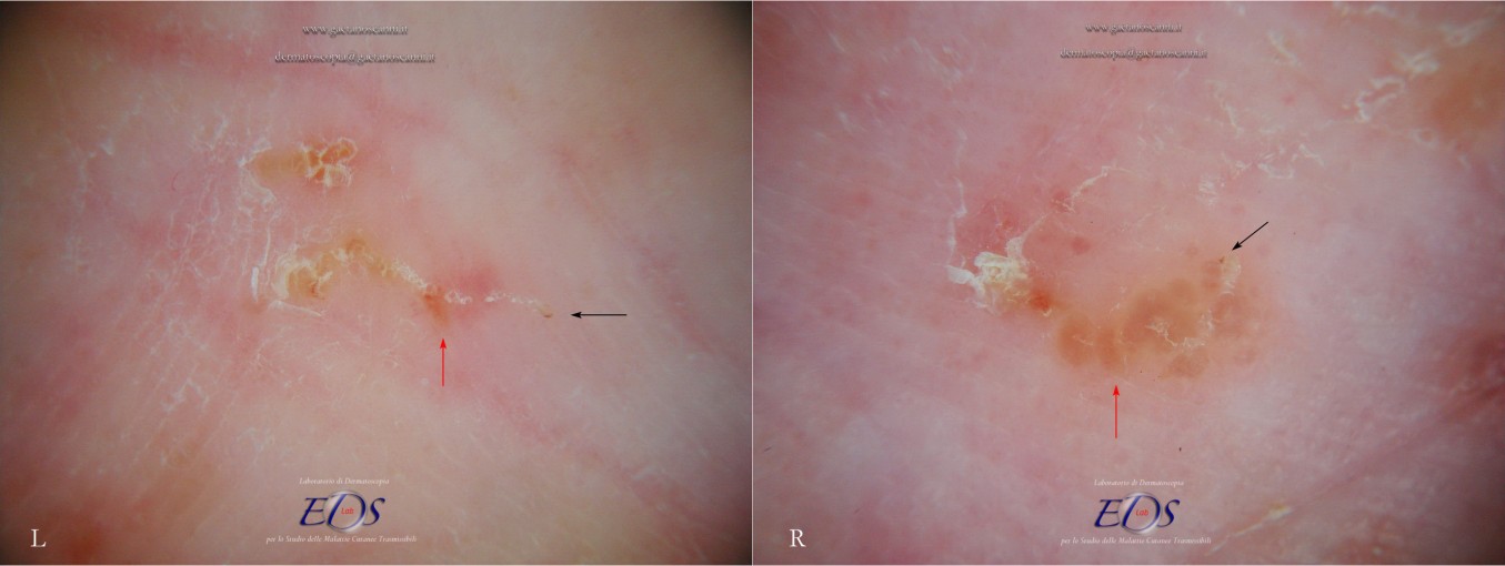
Fig 17. Phlogistic response to an MGU observed with polarized light contactless dermoscopy (d-DS). Next to the mite (black arrows, L-R) the skin is normal, while in the central part of the gallery an erythematous halo (red arrow, L) is evident. Immediately behind the mite, phlogosis can also occur in micro-vesicles trapped in the epidermis (red arrow, R). The tail of the galleries is characterized by keratinic collarettes.
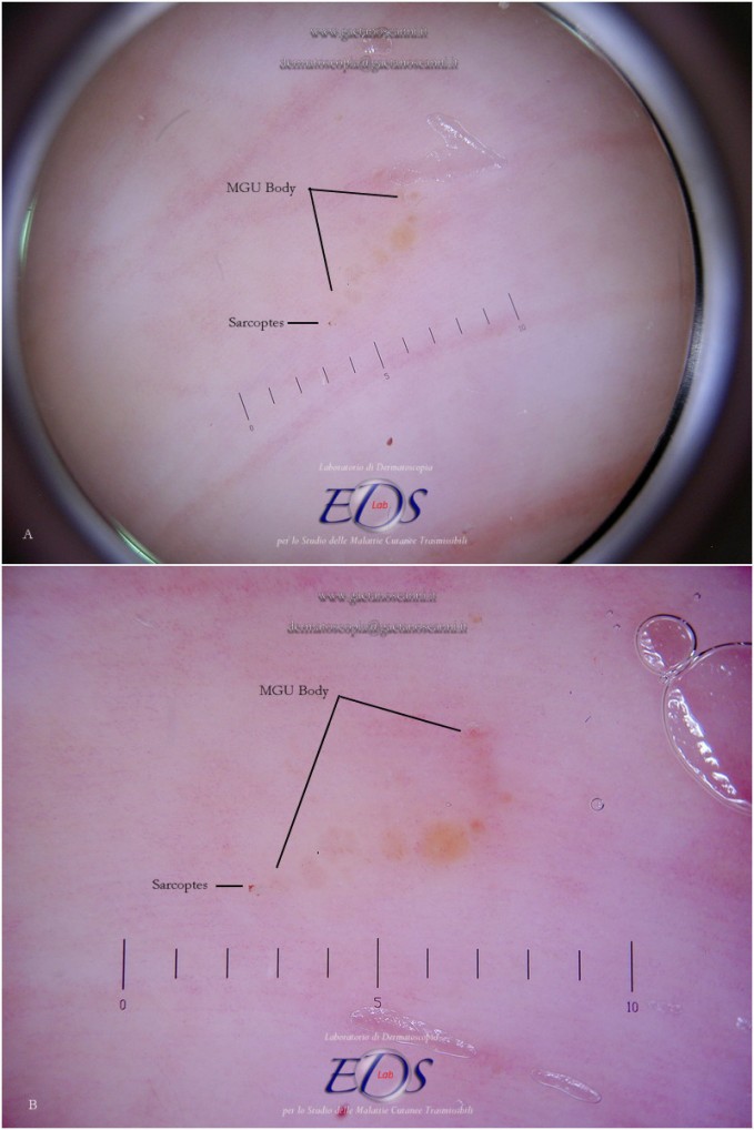
Fig 18. Contact dermoscopy (w-DS). The inflammatory response is an exudation that can remain trapped in skin thickness as a dyshidrotic-like appearance. In these images, the bubbles of MGU gallery were intentionally eliminated by moving the dermatoscope in order to better observe the phlogistic phenomenon.
04 MGU NODULES
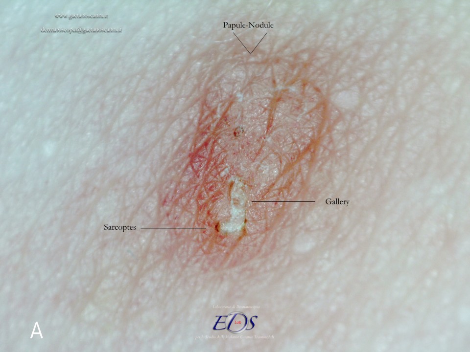
Fig 19. Enhanced contactless dermatoscopy (d-DS). Mite and gallery are clearly recognized in a recent papulo-nodular lesion.
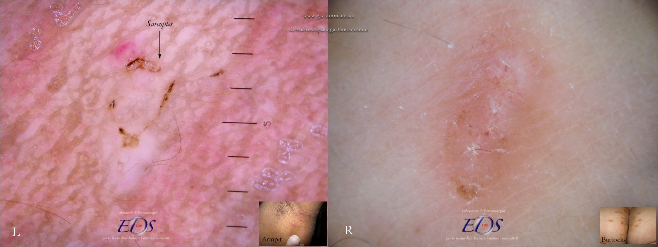
Fig 20. Nodular scabies observed in contact dermoscopy (w-DS) and contactless (d-DS) polarized light dermoscopy. A mite is recognizable in one of the axillary (L inset) papules of a subject of African ethnicity. A buttock (R inset) nodule present for a few weeks appears uninhabited instead.
05 MGU IMMATURE FORM
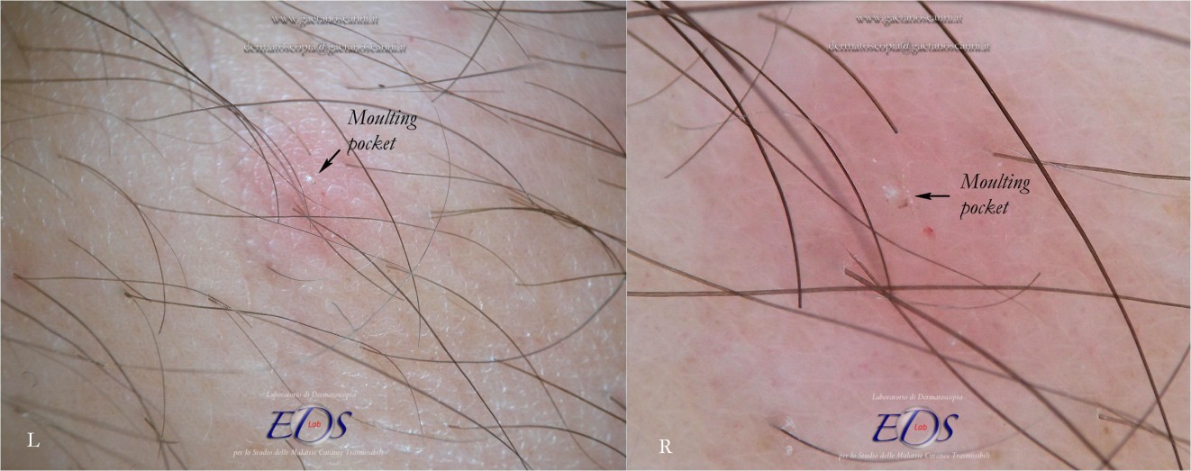
Fig 21. Moulting pocket in contactless dermoscopy (d-DS) with non-polarized (L) and polarized light (R). In this structure the complete Mite Gallery Unit does not develop because the mite remains stationary until the moulting process is completed or mating happens. There is instead only a small whitish raising (arrows, L-R) corresponding to the whole body of theSarcoptes behind which a real tunnel is not formed. Around the pocket, an area of erythema with blurred borders is recognizable.
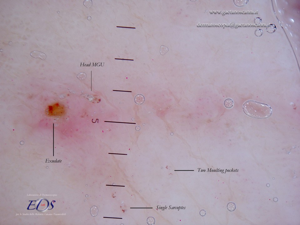
Fig 22. Contact dermatoscopy (w-DS). Some moult pockets are found in proximity to an adult head MGU which size is about twice larger than immature forms.
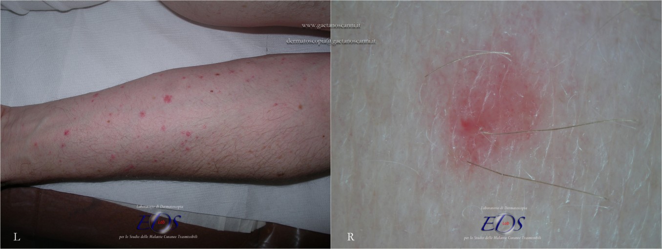
Fig 23. Scabies pseudo-follicular eruption observed under contactless dermoscopy (d-DS). Folliculitis-like lesions (L) are commonly considered non-specific sign of scabies. Under dermoscopy, only a peri-follicular erythematous halo is appreciated with some pinpoints-like vessels inside it (R). There are no signs of any complete or atypical MGU.
06 MGU GHOST
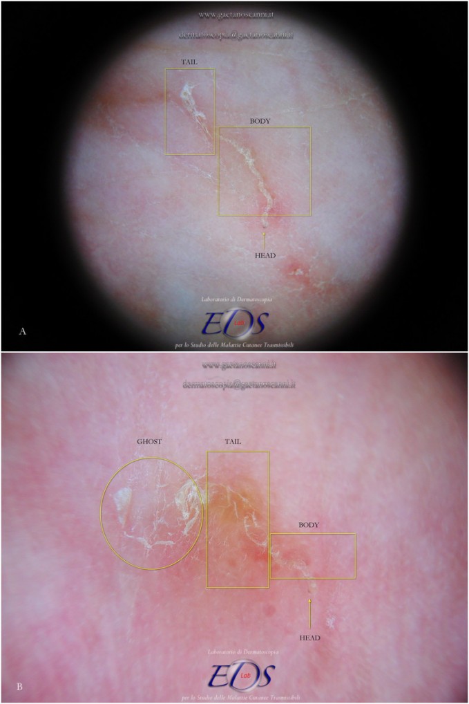
Fig 24. Contactless dermoscopy (d-Ds). In these pictures it is possible to follow the natural evolution of the oldest part of the MGU. (A) The tail of the MGU is constituted by a gallery which progressively loses the roof and of which only its bases remain as two keratinic edges. Subsequently they diverge to become areas of skin limited by thin polycyclic keratin flaps (B) in which the gallery is no longer recognizable (ghost gallery).
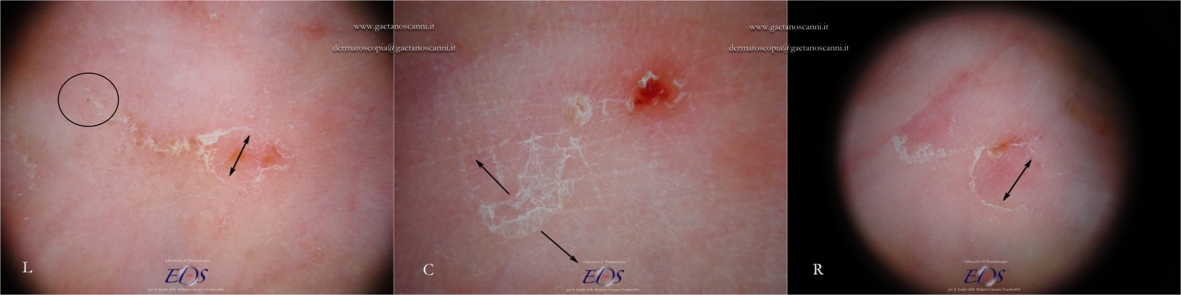
Fig 25. Evolution of an MGU towards a "ghost gallery" under polarized contactless dermoscopy (d-DS). A mature MGU ends with a tail made up of keratinic collarettes that progressively move away from each other (black arrow, L). Sarcoptes is identifiable in the front part of burrow (circle, L). When the mite is at the end of its life cycle or after therapy also the other parts of the tunnel undergo the normal processes of rearrangement of the skin (C) becoming thin and polycyclic keratinic edges of a ghost gallery (C,R).
Fine